
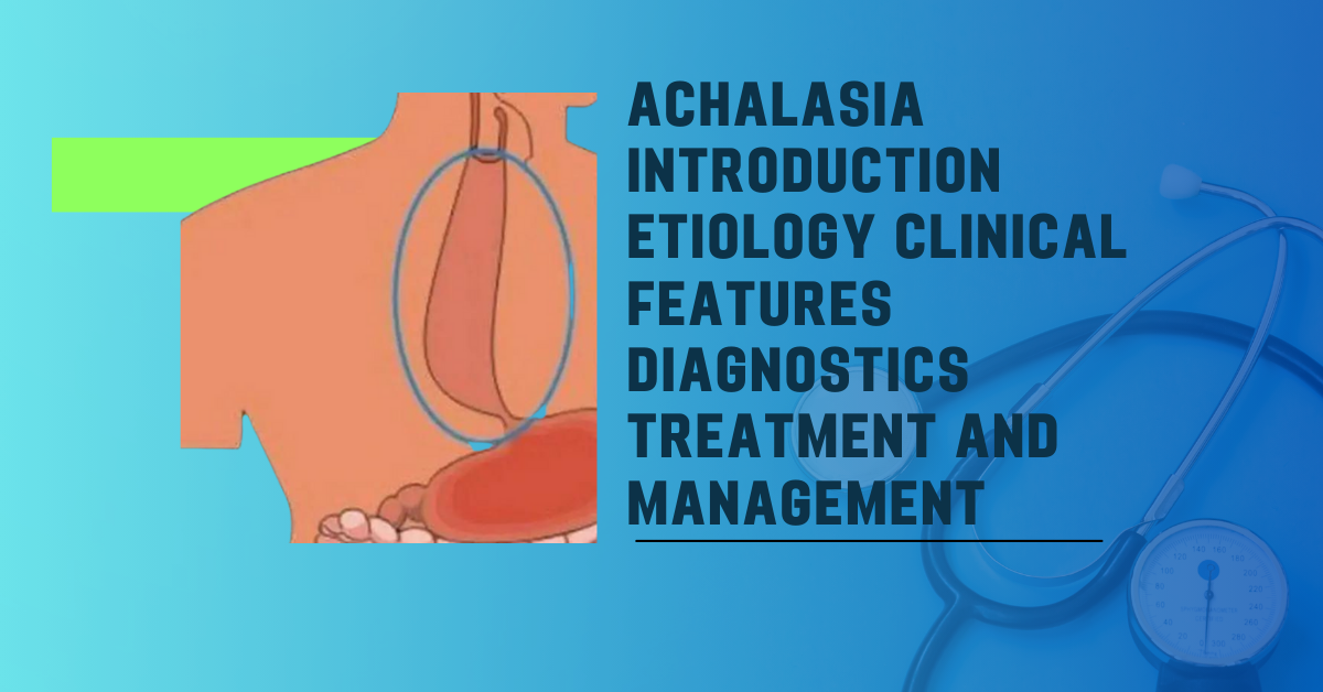
Achalasia is defined as the failure of the lower esophageal sphincter to relax during swallowing and non-peristaltic contractions in the distal part of the esophagus.
Achalasia Definition:
- Achalasia is an esophageal motility disorder characterized by the failure of the lower esophageal sphincter (LES) to relax during swallowing and non-peristaltic contractions in the distal two-thirds of the esophagus.
- It results from the degeneration of inhibitory neurons in the esophageal wall.
Etiology:
- Primary Achalasia: Often idiopathic and the most common cause.
- Secondary Achalasia (Pseudoachalasia): Occurs due to mechanical obstruction, mimicking achalasia. Underlying causes can include esophageal cancer, stomach cancer, Chagas disease, sarcoidosis, and amyloidosis.
Pathophysiology
- Swallowing regulation involves excitatory (Substance P, acetylcholine) and inhibitory (VIP, NO) neurotransmitters.
- In Achalasia, inhibitory neurons in the Auerbach (myenteric) plexus degenerate, resulting in a lack of inhibitory (VIP, NO) neurotransmitters.
- This leads to the inability of the LES to relax and an increased resting pressure of the LES, causing esophageal dilation proximal to the LES.
Clinical Features
Achalasia is characterized by the following clinical features:
- Dysphagia to both solids and liquids.
- Dysphagia can be progressive or paradoxical.
- Retrosternal pain.
- Regurgitation.
- Weight loss.
- Nocturnal cough.
Diagnostics
Diagnostic Approach:
- After clinical suspicion, patients should undergo an initial evaluation with:
- Esophageal barium swallow.
- Upper endoscopy to facilitate initial diagnosis.
- Regardless of initial findings, esophageal manometry is indicated to confirm the diagnosis.
Diagnostic Tests:
- Esophageal Barium Swallow:
- Best initial test.
- Often shows a “Bird’s beak” appearance: dilation of the proximal esophagus and narrowing of the gastro-esophageal junction, as well as delayed barium emptying or retention.
- Upper Endoscopy:
- To rule out malignancy or pseudoachalasia.
- May be normal in achalasia but may show retained food in the esophagus. Biopsy is indicated if malignancy is suspected.
- Esophageal Manometry:
- Confirmatory test.
- Typical findings include high resting pressure in the LES and absent peristalsis or uncoordinated contractions in the lower two-thirds of the esophagus.
Treatment
The choice of treatment depends on the patient’s surgical risk:
- High Surgical Risk:
- Endoscopic injection of botulinum toxin into the LES.
- Suitable for poor surgical candidates but may require repeated injections (every 6-12 months).
- Calcium channel blockers and nitrates are alternative options.
- Low Surgical Risk:
- Pneumatic dilation (endoscopic-guided balloon dilatation): Tears esophageal muscle fibers by dilating the esophagus with a balloon.
- Heller’s myotomy: Involves a longitudinal incision of the lower esophageal sphincter to facilitate food passage.
Complications
Complications of achalasia include:
- Squamous cell carcinoma (occurs in 5% of cases and is a serious concern).
- Aspiration pneumonia.
- Airway obstruction.
Key Point: It is essential to exclude esophageal carcinoma at the gastro-esophageal junction before initiating treatment for achalasia, as the symptoms can mimic achalasia. This is typically done through esophageal endoscopy.